

|
research |

|
G |
|
N |
|
I |
|
R |
|
E |
|
E |
|
N |
|
I |
|
G |
|
E |
|
U |
|
S |
|
S |
|
I |
|
T |
|
B |
|
A |
|
L |
|
A |
|
I |
|
R |
|
E |
|
T |
|
A |
|
M |
|
& |
|
S |
|
L |
|
E |
|
N |
|
O |
|
R |
|
A |
|
T |
|
O |
|
R |
|
Y |
|
B |
|
I |
|
O |

|
& |
|
T E B L |

|
& |
|
Tissue Engineering Biomaterials Laboratory |
|
University of Maryland Fischell Department of Bioengineering 3238 Jeong H. Kim Engineering Building College Park, Maryland 20742 P: 301.405.7475 F: 301.314.6868
© Copyright 2012 Tissue Engineering & Biomaterials Laboratory, TEBL, All Rights Reserved
|


perfusion bioreactors |
|
|
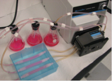
In vitro and in vivo nutrient transfer limits must be overcome in order to increase the feasibility of cell based therapeutic strategies. To enhance in vitro nutrient transport, the tubular perfusion system (TPS), a novel bioreactor recently developed by our laboratory, will dynamically culture human mesenchymal stem cells (hMSCs) in three dimensional scaffolds. This system utilizes an elegant design to create an effective cell culture environment without the drawbacks often associated with more complicated perfusion systems. The TPS design consists of hMSCs encapsulated in alginate beads which are tightly packed in a tubular growth chamber. Perfusing media through this growth chamber enhances nutrient transfer while exposing the cells to shear stress. To enhance in vivo vascularization, a prevascular network will be templated within the engineered tissue prior to implantation. To accomplish this, the TPS bioreactor will be optimized to support a coculture of endothelial cells and hMSCs. This strategy allows for the in vitro culture of functional engineered tissue, provides an elegant method for the in vivo implantation of the tissue, and fosters rapid integration of the implanted tissue into the host vasculature. Successful completion of these studies will demonstrate the feasibility of this fundamental technology for enhanced in vitro and in vivo nutrient transfer within cell based devices. |
|
|
vascular grafts |
|
|
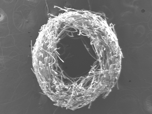
Coronary artery disease is a leading cause of mortality in both developed and developing nations. To treat diseased arteries, permanent metal stents are implanted in vessels in an attempt to restore bloodflow through the arteries. These stents remain permanently implanted and may cause both short-term and long-term complications. In other instances, it is necessary to bypass the occluded vessels and autologous vessels can be transplanted from a patient’s own vasculature to replace the blocked artery. Autologous vessels, however, may be unavailable due to a variety of reasons and surgeons often use synthetic grafts as an alternative. While permanent, synthetic vascular grafts have been employed successfully in large-diameter applications (>6mm), small-diameter grafts are plagued by restenosis and thrombosis, rendering them ineffective. Tissue-engineering approaches to develop a biodegradable graft that promotes expedited, native tissue growth and repair offer a potential solution to these problems. We aim to develop a vascular graft utilizing biodegradable polymeric materials and functional molecules to provide a microenvironment that encourages vascular cell adhesion and proliferation. |
|
|
stem cells & cell adhesion |
|
|
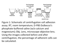
Mesenchymal stem cells (MSCs) are multipotent cells that have the ability to differentiate into various mesenchymal lineages such as bone, adipose, and cartilage. MSCs are typically harvested from the bone marrow and isolated based on their adherence to tissue culture plastic. There are several limitations to the traditional isolation method, including potentially harmful interactions between MSCs and the other cell types within the bone marrow, possible loss of late-adhering MSCs, and the undesired expansion of non-MSC adherent cells found in the bone marrow. In order to overcome the limitations of isolating MSCs, specific cell adhesion receptors, particularly integrins, may be targeted. By exploiting the interaction of integrins on the surface of MSCs with specific extracellular matrix ligands, it may be possible to isolate the MSC population from bone marrow aspirates with higher efficiency and specificity. Furthermore, adhesive interactions can be measured through the use of a cell adhesion assay in which cells are exposed to a surface for a period of time and then subjected to a known detachment force. Previously, we have used a modified centrifugation cell adhesion assay to study the relationship between chondrocyte adhesion and phenotype maintenance in 2D culture. Therefore, we propose to develop a modified polyethylene glycol (PEG) based material that will be capable of specific and efficient MSC isolation, while also directing MSC differentiation. The proposed PEG-based biomaterial will utilize specific ligands for integrin receptors that are present on the surface of MSCs.
|
|
|
craniofacial bone repair |
|
|
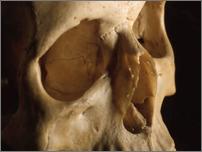
Orbital floor injuries are a devastating form of craniofacial trauma. Current clinical treatments, including the implantation of plastics or metals, are often inadequate as they may lead to a loss of function as well as poor aesthetics. Our strategy for orbital floor regeneration is based upon the in vitro preculture of osteoprogenitor cells within a degradable hydrogel. The proposed work investigates whether bone defects can be successfully treated by implanting an engineered bone graft that promotes osteoprogenitor cell differentiation by augmenting bone morphogenetic protein-2 expression, transport, and function. The successful completion of this work will allow us to develop a novel strategy for the treatment of orbital bone defects.
|
|
|
tebl home |
|
courses |
|
people |
|
research |
|
literature |
|
presentations |
|
journals |
|
links |
|
news |
|
tebl intranet |
|
|